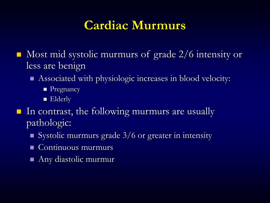

In dogs and cats, isolated diastolic murmurs are very rare. Murmurs may also be described as continuous that is, a murmur is continuously present throughout the cardiac cycle.

A diastolic murmur is heard during ventricular diastole that is, immediately after S2. Moderate murmur immediately audible with auscultationĪudible with stethoscope held slightly off chest wallĪ systolic murmur is one that is heard during ventricular systole that is, between the first (S1) and second (S2) heart sounds. Soft murmur that is audible with careful auscultation

Very soft murmur that is not immediately audible but can be heard only after careful auscultation in a quiet environment For example, a dynamic grade I–III/VI murmur describes a murmur that has an intensity that varies throughout the auscultation period, from difficult to hear (grade I) to immediately identifiable (grade III). 1 If murmur grade varies, as it often does in cats, the term "dynamic" is used and the grade is given as a range. Murmur intensity or "grade" is defined below. Intensity refers to the loudness of a murmur. When attributable to cardiac disease, murmur characteristics reflect which underlying cardiac disease may be present.ġ. Accurate description of a heart murmur facilitates determination of its possible genesis, whether it be physiologic or attributable to cardiac disease. Heart murmurs are routinely described by 1) intensity (loudness) 2) timing in the cardiac cycle and 3) location. In some cases, turbulent blood flow not only produces a heart murmur, but also a thrill that is palpable on the chest wall. When heart disease produces a heart murmur, it is because dynamic or fixed pathology disturbs laminar blood flow and creates turbulence. 1 In a normal heart, blood flow is characteristically laminar and hence silent.
#IVCD CARDIAC MURMURS SERIES#
Third and fourth heart sounds should not be appreciated in a normal dog or cat.Ī heart murmur is defined as a prolonged series of auditory vibrations emanating from the heart or blood vessels, most commonly attributable to turbulent blood flow. The second heart sound (S2) is audible at the completion of ventricular systole and is attributable to closure of the aortic and pulmonic valves. The first heart sound (S1) is attributable to closure of the mitral and tricuspid valves at the onset of systole. In a normal dog or cat, two heart sounds are audible. The second rhythm strip shows retrograde P waves just after the QRS complex.Physical examination of the cat or dog with suspected heart disease provides valuable information that allows "short-listing" of differential diagnoses to facilitate appropriate diagnostic and treatment recommendations. The strip below shows a junctional rhythm with retrograde P waves seen just before the QRS complex. AV blocking medications or electrolyte disturbances - is found. A pacemaker may be needed to relieve symptoms when no reversible cause - i.e. Often, the P wave is inverted in lead II, if it can be seen at all. The morphology of the P wave will not be similar to the sinus P wave, which is normally upright in lead II and biphasic in lead V1. When faster, it is referred to as an accelerated junctional rhythm.īecause the electrical activation originates at or near the AV node, the P wave is frequently not seen it can be buried within the QRS complex, slightly before the QRS complex or slightly after the QRS complex. A junctional rhythm is normally slow - less than 60 beats per minute. A junctional rhythm occurs when the electrical activation of the heart originates near or within the atrioventricular node, rather than from the sinoatrial node.īecause the normal ventricular conduction system (His-Purkinje) is used, the QRS complex is frequently narrow.


 0 kommentar(er)
